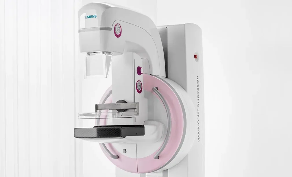
A mammogram is an X-ray of the breast. It uses X-rays to take images of the inside of the breast to detect any abnormalities. Mammography is best done during the time of the cycle when the breasts are least painful (the first part of the cycle). No preparation is needed before the mammogram.
On the day of the test, it is recommended that you do not use any cosmetic products (cream, cleansing milk, perfume, talcum powder) or wear any jewellery. You should also bring your old mammograms so that the radiologist can compare them with the new ones.
What is Mammography?
A mammogram is an X-ray image of the breasts taken with a low dose of X-rays. It is performed using a device called a mammograph, which may be equipped with a digital system for computerized image processing.
During a mammogram, radiological abnormalities are searched for, which could indicate breast cancer.
Mammography can be offered in several scenarios:
As part of an organised breast cancer screening campaign (biennial screening from 50 to 74 years of age); by your doctor for more frequent individual screening; when an abnormality is detected during a clinical breast examination; as part of a follow-up programme after breast cancer treatment. This test is carried out only with your consent. You are therefore free to accept or refuse it.
Mammograms in the Medical Imaging Department are performed with the latest generation of mammography machines, which makes the tests faster and with less radiation. Mammograms are performed with X-ray manipulators and analysed by a radiologist who can, if necessary, supplement the examination with a breast ultrasound in the ultrasound room next to the mammography room.
Frequently asked questions
Mammography is recommended in the first part of the menstrual cycle, i.e. before ovulation. The cycle usually lasts 28 days, so the optimal period would end on the 14th day after menstruation.
The radiology technician will place you correctly on the machine. Mammography is performed standing up with the patient naked. In order to obtain a good quality analysis and to see the whole breast, each breast is squeezed in turn between the two plates to spread the breasts out and avoid overlapping images. The sensation of breast compression is uncomfortable for some women, but only lasts a few seconds. A safety device limits the maximum pressure on the breast. Several images or incidences (usually 2 per breast) are taken from different angles to allow better viewing of the breasts. A basic mammogram takes 10-15 minutes on average.Afterwards, the radiologist examines the breasts and, if necessary, performs an ultrasound examination, which is completely painless and non-irritating. For young, pregnant or breastfeeding women, it is recommended that only the breast ultrasound is performed as a first-line procedure.
According to the National Public Health Agency, breast cancer accounts for one third of all new cancer cases in women and is the leading cause of cancer death in women. Although this pathology is sometimes detected at the onset of symptoms, the majority of women with breast cancer do not have any symptoms. That is why it is so important to have regular screening.

